Program details
Prevention of Transient Ischemic Attack (TIA)【Kishiwada Tokushukai Hospital】
surgery
Kishiwada Tokushukai Hospital(Kinki/Osaka)
Prevention of Transient Ischemic Attack (TIA)
Ensuring Safety Through Early Treatment
Transient ischemic attack (TIA) is a warning sign of cerebral infarction. Early detection and treatment of cervical and cerebral vascular stenosis are performed to prevent cerebral infarction through bypass surgery or stent placement.
- Genres
-
- Department
- Disease
- Examination Items/Treatments/Surgical method
- Region/Organ
- Program Summary
- Warning Signs of Stroke
Sudden weakness in one side of the body, slurred speech, or loss of vision in one eye that resolves within a few minutes.
These symptoms are warning signs of an impending stroke and are referred to as transient ischemic attacks (TIAs) in medical terms.
When atherosclerotic plaque (atheroma) accumulates inside the blood vessels of the neck (cervical vessels) or brain, causing the vessel lumen to narrow (a condition known as stenosis), blood flow is impeded. This reduces the blood supply to the brain, potentially leading to a stroke.
When blood stagnates near the site of stenosis, blood clots (thrombi) can form. If these clots dislodge and travel through the narrowed area to the blood vessels of the eye or brain, they can cause vision loss or stroke.
In some cases, the atheroma itself may rupture, and fragments of the plaque can travel to the brain's blood vessels, leading to a stroke.
Strokes caused by atheroma are referred to as atherothrombotic stroke.
Such stenosis commonly occurs in the cervical vessels and is known as carotid artery stenosis. This condition significantly increases the risk of future strokes and is considered a precursor to stroke, making it a critical issue.
Treatment is necessary before a stroke occurs.
In cases of mild stenosis, it is possible to manage the condition with medications that help thin the blood (antiplatelet agents). However, in cases of severe stenosis, surgical treatment is required.
Currently, there are two surgical options available. One involves performing a procedure under general anesthesia in an operating room. In this procedure, the cervical blood vessels are exposed, and the affected vessel is incised to remove the plaque adhered to the inner wall. This surgery is known as carotid endarterectomy.
The other method, performed under local anesthesia in a catheterization room, involves using a balloon catheter to widen the narrowed area from inside the blood vessel. A mesh-like metal tube called a stent is then placed to push the plaque against the vessel wall, keeping the vessel open. This procedure, known as carotid artery stenting, is a type of endovascular treatment.
Unlike the first method, this approach leaves no scars on the neck and typically requires only a 4–5 day hospital stay.
Nowadays, extremely thin stents designed for use in cerebral blood vessels are also available.
When a blood vessel is narrowed, the affected area can be widened using a balloon or stent. However, if the blood vessel is already completely blocked, it cannot be reopened. This condition is referred to as occlusion.
Even if a blood vessel is occluded, some individuals remain asymptomatic and live without realizing it, as their brain naturally develops collateral circulation (bypass pathways).
However, if dehydration occurs, causing blood to become concentrated, or if blood pressure drops too low, the collateral pathways may not be sufficient to deliver blood to all areas of the brain. In such cases, transient ischemic attacks (TIAs) or strokes may occur.
When this happens, it may be necessary to create an artificial bypass to restore adequate blood flow to the affected areas of the brain.
In general, a bypass surgery is performed by connecting a blood vessel located under the skin in the temple area (the superficial temporal artery) to a blood vessel on the brain's surface (the middle cerebral artery).
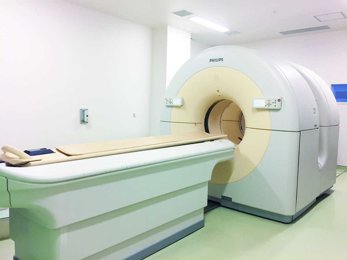
Narrowing or occlusion of the cervical or cerebral blood vessels can be easily examined in outpatient settings using MRI scans designed to visualize blood vessels in the brain and neck, known as MRA, or carotid artery ultrasound.
If you have been told during a brain checkup or detailed examination that your brain or neck blood vessels are narrow or blocked, please consult us.
- Medical Institutions
-
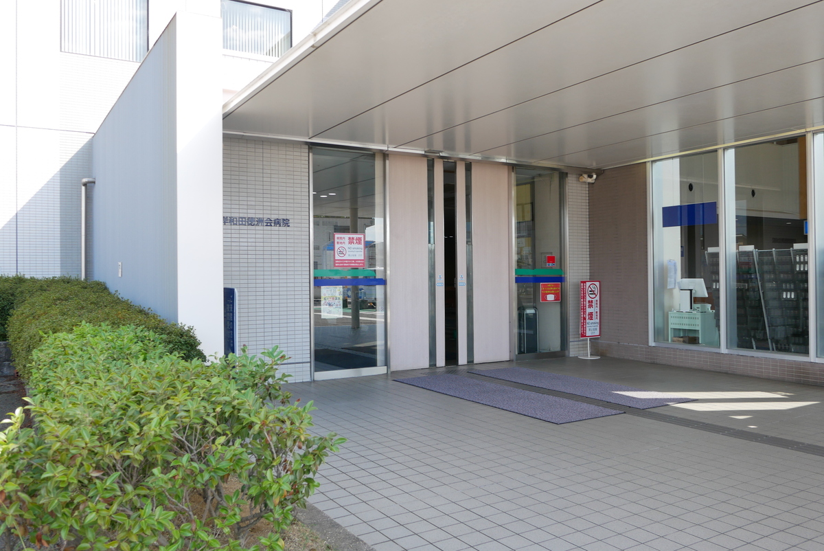
Kishiwada Tokushukai Hospital
〒596-0042
4-27-1 Kamoricho, Kishiwada City
- Examination Items
- Setup Date
- Excluded days
- Required Days/Hours
- Start/end time
- Eligibility Criteria/Exclusions for Treatment
- Admission Criteria
1. Medical Information:
- Diagnosis (e.g., transient ischemic attack, carotid artery stenosis, cerebral vascular stenosis)
- Detailed symptoms (e.g., weakness in limbs, speech impairment, vision disturbances)
- Examination results from other institutions (e.g., MRI, MRA, carotid ultrasound)
- Presence of comorbidities (e.g., hypertension, diabetes, heart disease)
2. History of Treatment:
- History of cerebrovascular treatments (e.g., bypass surgery, stent placement)
- Current medications (e.g., anticoagulants, antiplatelet agents)
- Presence of allergies or adverse drug reactions
3. Age and Physical Fitness:
- Evaluation of whether elderly patients or those with comorbidities can withstand surgery or treatment
- Suitability for general or local anesthesia
4. Renal Function:
- Assessment of renal function is required due to potential use of contrast agents
5. Severity and Urgency of Symptoms:
- Frequency and timing of transient ischemic attacks or cerebral vascular stenosis
- Assessment of whether there is a high risk of cerebral infarction and whether urgent intervention is needed
6. Other Conditions:
- Ability to follow treatment instructions for blood pressure management and thrombosis prevention
- Willingness and ability to cooperate in lifestyle improvement and follow-up monitoring
- Precautions / Contraindications
- 【Precautions and Contraindications】
1. Cases Not Suitable for Treatment:
- In cases of impaired renal function, the use of contrast agents may not be feasible, limiting catheter-based treatment.
- Difficulty in discontinuing antiplatelet or anticoagulant medications increases the risk of bleeding and may restrict treatment options.
- Severe cardiopulmonary dysfunction or insufficient physical strength to tolerate surgery may also render treatment unsuitable.
2. Contraindications Related to General or Local Anesthesia:
- Patients with comorbidities that complicate the management of breathing or circulation during anesthesia require careful consideration of anesthesia methods.
- A history of allergies or hypersensitivity reactions to medications necessitates prior confirmation of the drugs to be used.
3. Treatment Risks and Complications:
- Surgical or catheter-based treatment carries risks of complications such as bleeding, thrombus formation, and cerebral infarction, with intracranial complications potentially having severe consequences.
- Risks such as arterial dissection or vascular rupture may occur, and regular postoperative follow-up is crucial.
---
# 【Important Pre-Treatment Information】
1. Preoperative Preparation:
- Follow medical instructions regarding the discontinuation of antiplatelet or anticoagulant medications and ensure proper medication management before treatment.
- Preoperative tests (e.g., blood tests, MRI, ultrasound) are essential for accurate diagnosis and treatment planning.
2. Postoperative Care and Follow-Up:
- Monitor for risks such as bleeding, thrombus formation, or infection, and contact a medical institution immediately if abnormalities occur.
- Follow-up after bypass surgery or stent placement is necessary, and adhering to postoperative instructions and regular visits is crucial.
3. Lifestyle and Rehabilitation:
- Postoperative adjustments to exercise and lifestyle habits are recommended, with activities guided by medical advice.
- Focus on cerebrovascular health through smoking cessation, a balanced diet, and moderate exercise.
4. Hospitalization and Follow-Up Schedule:
- Confirm the length of hospitalization and rehabilitation period in advance and plan accordingly.
- Continue regular follow-up visits after discharge to monitor progress appropriately.
5. Emergency Response:
- If severe headache, vision impairment, or limb paralysis occurs after discharge, contact a medical institution immediately.
- Confirm the contact details of a medical facility or physician available for emergencies in advance.


