Facility Details
View an overview of the medical facility, affiliated physicians, and available programs in one place.
- Kinki / Osaka
- medical treatment
- regenerative medicine
- orthopaedic surgery
- health check up
- stem cell therapy
- arthrosis
- MRI/MRA
- brain
- DWIBS
- gynecology
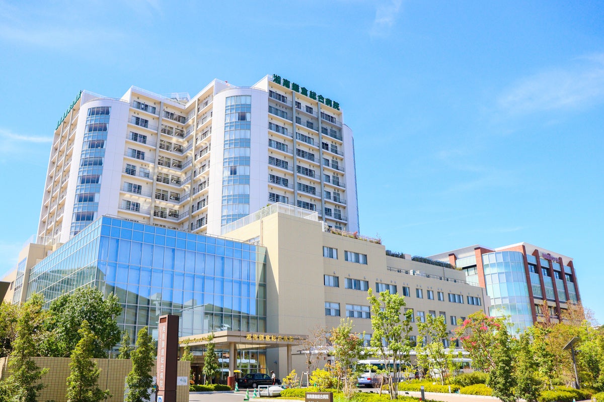
SOBAJIMA CLINIC
Sobajima clinic specializes in osteoarthritis treatment with cell transplantation and is one of the country’s best regenerative medicine clinic. Well experienced doctors and stuffs offer safe and body-friendly treatment in our clinic.
Sobajima Clinic is a regenerative medicine clinic with one of the leading track records in Japan for the treatment of osteoarthritis using cell transplantation.
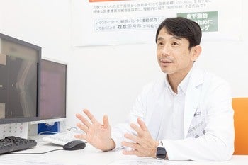
The director has consistently pursued research in regenerative medicine since studying in the United States, aiming to provide treatment options that do not rely on surgery. Through collaborative research with the Department of Orthopedics at Kobe University, the clinic obtained approval for regenerative medicine, and in November 2016 performed its first case for knee osteoarthritis.
Over nearly a decade, the clinic has accumulated extensive clinical experience. Today, with certified regenerative medicine specialists, highly trained staff, and advanced equipment, the clinic provides treatments with a strong focus on safety and effectiveness.
The clinic is also actively engaged in medical research to improve treatment quality and has published 10 academic papers to date.
With the goal of extending patients’ healthy lifespan, the clinic emphasizes not only single treatments but also continuous support, including multiple treatment sessions and rehabilitation.
- Location
- 〒577-0011
Clinic court Higashino, 2-2-6 Aramoto-kita, Higashi Osaka City Osaka
- Website
- https://sobacli-saisei.com/
- Specialty
- 【Departments / Services】
Orthopedics (General)
Sports Orthopedics
Regenerative Medicine (Osteoarthritis)
Stroke & Lifestyle-related Diseases
Dementia
Rehabilitation
Breast Clinic
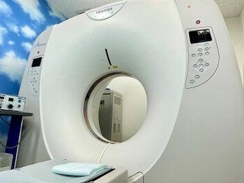
Various MRI Examinations
【Health Checkups】
Whole-body MRI Cancer Screening (DWIBS)
Painless Breast MRI Screening (DWIBS)
Brain Checkup (Head MRI & MRA)
Brain Checkup (Head MRI & MRA + Neck MRA)
Head & Cervical Spine Checkup
Breast Cancer Screening (Mammography + Breast Ultrasound + Physical Examination + Consultation)
Spine Checkup (Cervical & Lumbar MRI)
Osteoporosis Checkup (Thoracic & Lumbar MRI + Bone Density Test)
Brain Checkup with AI-based Dementia Risk Assessment
【Treatments】
PRP Therapy for Orthopedic Conditions
Extracorporeal Shockwave Therapy
Stem Cell Therapy (Orthopedic Conditions)
Stem Cell Therapy for Chronic Pain (e.g., numbness from spinal canal stenosis)
Stem Cell Therapy for Post-stroke Sequelae
- Facility details
- 【Location】
Located along Osaka Chuo-Odori Avenue
1-minute walk from Aramoto Station (Osaka Metro Chuo Line)
Near the intersection of Hanshin Expressway Higashi-Osaka Line and Kinki Expressway
【Equipment】
1.5T MRI (GE Healthcare)
Focused Extracorporeal Shockwave Therapy System (Storz)
Ultrasound Devices
Mammography System
X-ray Equipment
Body Composition Analyzer
【Facilities】
On-site Cell Processing Center (CPC)
Rehabilitation Facilities
Operating Room
【Others】
• Hotel New Otani Osaka (6th Floor)
Special discounted accommodation rates are available for our patients.
• Adipose-derived stem cells have various effects such as anti-inflammatory action, angiogenesis, and tissue protection.
By injecting a patient’s own stem cells into joints affected by osteoarthritis (such as knees and hips), pain relief and functional recovery can be expected.
We also provide stem cell therapy for chronic pain (e.g., neuropathic pain caused by spinal canal stenosis) and for post-stroke sequelae.
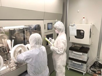
• Our clinic has an in-house Cell Processing Center (CPC).
With 20 years of basic research, we utilize optimized cell extraction and culture techniques to safely process and provide high-quality cells.
https://soba-cli.com/
Available Programs
-
Combination Therapy Using Two Types of Adipose-Derived Stem Cells (Fresh Cells + Cultured Cell Aggregates) [Sobajima Clinic]
Combination Therapy Using Fresh Cells and Cultured (Spheroid) Cells
1.Fresh Cells
(Freshly harvested stem cells)
2.Cultured Spheroid Cells
(Culture-expanded spheroid stem cells)Medical Treatment: Two Types of Stem Cell Therapy 1.Fresh Cell Therapy Fresh stem cells are extracted from approximately 100 cc of adipose tissue harvested under local anesthesia and administered to the affected area on the same day (outpatient procedure). 2.Cultured Spheroid Cell Therapy A portion of the fresh cells is cryopreserved and cultured in our in-house Cell Processing Center (CPC) to form spheroids. Approximately one month after the start of culture, the spheroid cells are administered to the affected area. After the initial fresh cell injection, additional treatments are provided periodically based on the patient’s condition. Treatment Features • Fresh cells consist of a heterogeneous population of cells with diverse functions, including anti-inflammatory effects, angiogenesis, and tissue repair. Each cell type plays a specialized role. • Subsequent treatment using spheroid cell aggregates is considered to provide greater therapeutic effects compared to administering individual dispersed cells. • This combination therapy approach first suppresses inflammation, promotes blood vessel formation, and prepares the tissue environment for repair. Following this, spheroid cell therapy further optimizes the environment and enhances tissue regeneration. Indications / Conditions 1.Osteoarthritis 2.Rheumatoid Arthritis (refractory cases) 3.Injuries of muscles, tendons, ligaments, and other periarticular tissues
View Details -
Adipose-Derived Stem Cell Therapy (Fresh Cells and Cultured Cells) [Sobajima Clinic]
In addition to cultured cells, fresh cells are administered either via local injection or intravenous infusion.
1.Fresh Cells
(Fresh Cells: Freshly harvested stem cells)
2.Cultured Cells
(Cultured Cells: Culture-expanded stem cells)Medical Treatment: Two Types of Stem Cell Therapy 1.Fresh Cell Therapy Fresh stem cells are extracted from approximately 100 cc of adipose tissue harvested under local anesthesia. These cells are administered on the same day either: by local injection into the affected area (e.g., joints), or via intravenous infusion (IV drip). This procedure is performed on an outpatient basis (same-day surgery). 2.Cultured Cell Therapy A portion of the fresh cells is cryopreserved and cultured in our in-house Cell Processing Center (CPC). Approximately one month after the start of culture, the cultured cells are administered either: by local injection into the affected area (e.g., joints), or via intravenous infusion. Following the initial fresh cell treatment, cultured cell therapy is provided periodically based on the patient’s clinical condition. Indications / Conditions 1.Osteoarthritis 2.Rheumatoid arthritis (refractory cases) 3.Residual numbness or pain in the upper or lower limbs due to spinal disorders or cerebrovascular diseases (including persistent symptoms after surgery) 4.Chronic pain remaining after standard treatments or surgical interventions 5.Injuries of muscles, tendons, ligaments, and other periarticular tissues
View Details -
Whole-body MRI Cancer Screening (DWIBS) [Sobajima Clinic]
DWIBS is an advanced screening method that uses MRI to evaluate cancer risk throughout the entire body.
Because it does not use radiation, it places less burden on the body.Features • DWIBS is an advanced screening method that uses MRI to evaluate cancer risk throughout the entire body. • It does not require dietary restrictions, contrast agents, or radiation exposure, resulting in minimal burden on the body. • In approximately 30–40 minutes, imaging from the neck to the pelvis can be performed in a single session, contributing to early cancer detection.
View Details -
Non-invasive Breast MRI Cancer Screening (DWIBS) [Sobajima Clinic]
This MRI examination can be performed comfortably while wearing examination clothing, without pain.
As it does not use radiation, it places minimal burden on the body.Features • No breast compression, so the examination is painless • Performed while wearing clothing, ensuring privacy • High cancer detection rate (no blind spots, high accuracy) • No radiation exposure, ensuring safety • Reliable post-examination follow-up by breast specialists
View Details -
Breast Cancer Screening with Same-Day Results (Mammography & Ultrasound) [Sobajima Clinic]
This is a comprehensive screening plan that allows breast cancer examination and results explanation to be completed in a single day.
Features • A comprehensive screening plan that completes breast cancer examination and results explanation in one day ※ The breast specialist is a male physician who also serves as a specially appointed professor at a university ※ Mammography is performed by a female technician
View Details -
Brain Health Screening (Head MRI/MRA and Neck MRA) [Sobajima Clinic]
MRI and MRA of the head, along with MRA of the neck, are used to comprehensively evaluate the brain and blood vessels, allowing detailed assessment for conditions such as cerebral infarction, aneurysms, and arteriosclerosis.
Recommended For • Individuals concerned about brain diseases such as stroke • Those with a family history of stroke • Individuals who wish to undergo regular brain checkups • Those concerned about arteriosclerosis Comprehensive MRI Examination from Brain to Neck • A 1.5 Tesla MRI is used to perform diagnostic imaging of the entire head region, from the brain to the neck. • Head MRI provides cross-sectional images of the intracranial area to detect signs of conditions such as cerebral infarction, brain tumors, and encephalitis. Head MRA creates detailed three-dimensional images of the cerebral blood vessels, enabling evaluation of vascular narrowing, blood flow abnormalities caused by infarction, subarachnoid hemorrhage, and cerebral aneurysms. • Neck (cervical) MRA generates detailed three-dimensional images of the cervical blood vessels and assesses the degree of arteriosclerosis in the carotid arteries, which is a major risk factor for stroke.
View Details -
Brain Health Screening (Head MRI/MRA ) [Sobajima Clinic]
MRI and MRA of the head are used to comprehensively evaluate the brain and blood vessels, allowing detailed assessment for conditions such as cerebral infarction, aneurysms, and arteriosclerosis.
Recommended For • Individuals concerned about brain diseases such as stroke • Those with a family history of stroke • Individuals who wish to undergo regular brain checkups MRI Examination of the Entire Head • A 1.5 Tesla MRI is used to perform diagnostic imaging of the entire head. • Head MRI provides cross-sectional images of the intracranial area to detect signs of conditions such as cerebral infarction, brain tumors, and encephalitis. Head MRA creates detailed three-dimensional images of the cerebral blood vessels, enabling evaluation of vascular narrowing, blood flow abnormalities caused by infarction, subarachnoid hemorrhage, and cerebral aneurysms.
View Details -
Brain Screening with AI-Based Dementia Prevention Assessment[Sobajima Clinic]
MRI and MRA of the head, along with MRA of the neck, are used to comprehensively evaluate the brain and blood vessels, enabling detailed assessment of conditions such as cerebral infarction, aneurysms, and arteriosclerosis.
In addition, this examination utilizes AI analysis (BrainSuite™), supervised by the Institute of Development, Aging and Cancer at Tohoku University, to assess the risk of dementia.Recommended For • Individuals who have never undergone a brain checkup • Those who wish to have regular brain screenings • Individuals concerned about Alzheimer’s disease or dementia • Those who want early detection of brain disorders • Individuals who currently have no symptoms but feel concerned • Those worried about brain diseases such as stroke • Individuals with a family history of stroke MRI Examination of the Entire Head • A 1.5 Tesla MRI is used to perform diagnostic imaging of the entire head. • Head MRI provides cross-sectional images of the intracranial area to detect signs of conditions such as cerebral infarction, brain tumors, and encephalitis. Head MRA creates detailed three-dimensional images of the cerebral blood vessels, enabling evaluation of vascular narrowing, blood flow abnormalities caused by infarction, subarachnoid hemorrhage, and cerebral aneurysms. What is AI-based Dementia Prevention Assessment? • Conventional brain checkups alone make it difficult to accurately assess dementia risk and provide effective prevention. • “BrainSuite™,” supervised by the Institute of Development, Aging and Cancer at Tohoku University, analyzes head MRI images using artificial intelligence (AI). Based on advanced brain research, it evaluates your brain health condition. • Furthermore, it provides personalized advice to help maintain and improve brain health, supporting the creation of a “healthy brain” aimed at preventing dementia.
View Details -
Comprehensive Head and Cervical Spine MRI Screening[Sobajima Clinic]
MRI and MRA of the head are used to comprehensively evaluate the brain and blood vessels, allowing detailed assessment of conditions such as cerebral infarction, aneurysms, and arteriosclerosis.
This examination is also recommended for individuals experiencing neck or shoulder stiffness.Recommended For • Individuals concerned about brain diseases such as stroke • Those with a family history of stroke • Individuals who wish to undergo regular brain checkups • Those concerned about arteriosclerosis Comprehensive MRI Examination from Brain to Cervical Spine • A 1.5 Tesla MRI is used to perform diagnostic imaging of the entire region from the brain to the cervical spine. • Head MRI provides cross-sectional images of the intracranial area to detect signs of conditions such as cerebral infarction, brain tumors, and encephalitis. Head MRA creates detailed three-dimensional images of cerebral blood vessels, enabling evaluation of vascular narrowing, blood flow abnormalities caused by infarction, subarachnoid hemorrhage, and cerebral aneurysms. • Cervical spine MRI evaluates conditions such as cervical tumors, spinal cord tumors, vascular tumors, and intervertebral disc herniation. • This examination is also recommended for individuals experiencing neck or shoulder stiffness.
View Details -
Osteoporosis Screening (Thoracic and Lumbar MRI with Bone Mineral Density Measurement) [Sobajima Clinic]
Bone density measurement is performed to evaluate bone health, and MRI allows detailed assessment of vertebral compression fractures (including old fractures), which are common osteoporotic fractures that may be difficult to detect on X-rays.
This examination is also recommended for individuals experiencing lower back pain.Recommended For • Postmenopausal women • Individuals experiencing lower back pain MRI Examination of Thoracic & Lumbar Spine with Bone Density Measurement • A 1.5 Tesla MRI is used to perform diagnostic imaging of the thoracic and lumbar spine. • Bone density measurement is conducted to evaluate bone health, and MRI enables detailed assessment of vertebral compression fractures (including old fractures), which are typical osteoporotic fractures that may be difficult to detect on X-rays. • As lumbar spine MRI is included, this course is also recommended for individuals concerned about lower back pain.
View Details -
Comprehensive Spine MRI Screening (Cervical and Lumbar Spine) [Sobajima Clinic]
Recommended for individuals experiencing shoulder stiffness or lower back pain.
This examination can evaluate not only bones but also the condition of nerves and intervertebral discs.Recommended For • Individuals experiencing shoulder stiffness or lower back pain MRI Examination of the Cervical and Lumbar Spine • A 1.5 Tesla MRI is used to perform diagnostic imaging of the cervical and lumbar spine. • This examination evaluates not only bones but also intervertebral discs and nerves, making it possible to identify the causes of persistent lower back pain, leg numbness, and shoulder stiffness. • It is useful for detecting conditions such as vertebral tumors, lumbar tumors, spinal tumors, intervertebral disc herniation, lumbar spinal canal stenosis, lumbar compression fractures (including old fractures), degenerative lumbar spondylosis, spondylolisthesis, and ossification of the posterior longitudinal ligament (designated intractable disease).
View Details